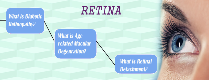

The retina is the light-sensitive layer of tissue that lines the inside of the eye and sends visual messages through the optic nerve to the brain. When the retina detaches, it is lifted or pulled from its normal position. If not promptly treated, retinal detachment can cause permanent vision loss. In some cases there may be small areas of the retina that are torn. These areas, called retinal tears or retinal breaks, can lead to retinal detachment.

Retinal Detachment

Rhegmatogenous Retinal Detachment
There are three different types of retinal detachment:
A retinal detachment can occur at any age, but it is more common in people over age 40. It affects men more than women, and Whites more than African Americans.
A retinal detachment is also more likely to occur in people who:
Symptoms include a sudden or gradual increase in either the number of floaters, which are little "cobwebs" or specks that float about in your field of vision, and/or light flashes in the eye. Another symptom is the appearance of a curtain over the field of vision. A retinal detachment is a medical emergency. Anyone experiencing the symptoms of a retinal detachment should see a retinal surgeon immediately.
Small holes and tears are treated with laser surgery or a freeze treatment called Cryopexy. These procedures are usually performed in the doctor's office. During laser surgery tiny burns are made around the hole to "weld" the retina back into place. Cryopexy freezes the area around the hole and helps reattach the retina.
Wet AMD happens when abnormal blood vessels behind the retina start to grow under the macula known as choroidal neovascular membrane (CNVM). These new blood vessels can be fragile and leak blood and fluid. The blood and fluid cause the macula to swell and damage occurs rapidly. The damage may also cause scarring of the retina.
In 90%- 95% patients retina gets attached and vision improves. But in 5 %- 10% patients develop re- detachment. In such cases resurgery is required. Resurgery success rate depends on retinal condition, amount of proliferative changes, and timing of resurgery. Success of retinal surgery is count on reattachment of retina and not on recovery of vision.
Visual recovery is depending on reattachment of retina and improvement in macular condition (central portion of retina). Recovery can take time from weeks to months and can be partial to full recovery. The main aim of treatment is to reattach retina into its place.
In retinal detachment treated patients after successful reattachment of retina every 3 month follow up for check up of same and fellow eye for at least one year. After that as per doctor’s suggestion follow up should be done.
© 2015 Drushti Eye Institute Pvt Ltd. All Rights Reserved
Developed by - Regal Soft India